Saturday, 22 September 2012
The 1001 Faces of the Neuron
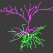
 The kind of diagram used to represent a typical neuron with its specialized extensions (axon and dendrites) can make us forget the unbelievable variety of shapes that these nerve cells can actuslly have. If you don’t believe it, go take a look at the web site NeuroMorpho.Org (click the link below).
This site contains a database of more than 6600 digitally reconstructed images of neurons and their complex branching structures, and even lets you view them in 3D. This image database was compiled from contributions from over 60 laboratories worldwide that use a variety of methods of staining and neuronal tracing in their experiments.
Most of the neurons depicted in this database come from the brains of mice, rats, and human beings, but some come from those of monkeys, cats, salamanders, flies, and even elephants—a total of 11 different species in all. Each image is accompanied by a generous amount of relevant data. You can search the database not only by species, but also by brain region , (cortex, hippocampus, etc.), cell type (pyramidal, motor neuron, etc.), and the name of the laboratory that provided the neuron images.
If you pass your mouse cursor over the names of the neurons, you will get visual overviews that give you an idea of their morphology. You can then click the name of the neuron that interests you and retrieve all of the information concerning it, plus a 3D animation in which it rotates so that you can appreciate it in all its beauty. A Java application also lets you manipulate each neuron image (zoom in, zoom out, etc.) and even download it for free.
NeuroMorpho.Org deserves a lot of credit for sharing all this knowledge free of charge—very much in the spirit of the Copyleft philosophy that guides The Brain from Top to Bottom .
From the Simple to the Complex | Comments Closed


