Monday, 25 November 2019
Three On-Line Atlases of the Human Brain

This week I want to tell you about three different on-line atlases of the human brain. All three will let you satisfy your curiosity by exploring the inmost recesses of the most complex object in the known universe, of which each of us has an example right between our ears. So stand warned: this post may well leave you spending even more time on your computer than usual!
First there is the Whole Brain Atlas, by Keith A. Johnson and J. Alex Becker,which has already been around for a while. On the home page, under the heading Normal Brain, the first topic is Normal Anatomy in 3-D with MRI/PET (Javascript). Here alone, you can have hours of fun clicking on the little black-on-yellow directional arrows to explore successive slices of the brain in the transaxial, sagittal and coronal planes. Often, you’ll see a large yellow arrow pointing at the specific brain structure whose name appears in the drop-down list box below the image, to the left. But you can also explore the other way around: click on the arrow in this list box to display a list of brain structures, then choose the one you’d like to view. An image of this structure then appears, with a big yellow arrow pointing at it.
Other topics in the Normal Brain section, such as Top 100 Brain Structures and Can you name these brain structures?, are designed to let you have fun while learning brain anatomy. The other main subject headings on the home page let you explore numerous images of abnormal brains, such as the brains of people who have had strokes or have brain tumours or various degenerative, inflammatory or infectious brain diseases.
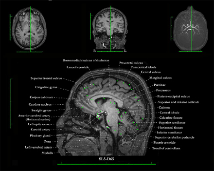
The second, similar atlas is the University of Chicago MRI Brain Atlas. The page How To Use the Interactive Interface explains the symbols and abbreviations used in this atlas and the various planes of the brain sections shown in it. To access the atlas, scroll down until you see the Start Atlas button at the centre of the page, then click it. You will then probably have to click one more time to agree for your browser to use Adobe Flash. Next you will land on the home page of the atlas, but you’ll still have a few steps to go. You must click the Start button to the lower right to choose which of the axial, coronal or sagittal sections you want to explore, and then choose between two major regions of the brain. Only then will you see a list of sections, with strange names such as CO35-COD50. Once you click the name of the section that you’re interested in, an image of it will appear at last. But the effort will have been worth it: the presentation is impeccable, and you can make the names of the visible brain structures appear one by one by clicking the lines pointing to them. Or you can make them all appear or disappear at once.
The third and last atlas that I want to mention is the Big Brain, published in June 2013, It is part of the Human Brain Project and was produced by an international team of neuroscientists from the Montreal Neurological Institute in Canada and the Forschungszentrum Jülich in Germany. These researchers sliced, scanned, and analyzed the brain of a deceased 65-year-old woman to create a detailed 3D computer model of an entire human brain. Thus, unlike the two other atlases, which consist of images of sections of living people’s brains, captured by magnetic resonance imaging (MRI), this one consists of coloured sections of a person’s brain post mortem. Compared with the MRI images, whose resolution is only 1 cubic millimetre, the images in the Big Brain were made from 7400 slices of the brain, each only 20-µm thick, thus enabling 50 times greater resolution. It took the team 1000 hours to image all of these slices with a scanner, thus generating the 1 billion billion bytes of data that they used to construct their 3D model. You can explore it by using your mouse wheel to zoom in and out on every image.
From the Simple to the Complex, Uncategorized | Comments Closed


