


Tuesday, 17 September 2024
Spectacular reconstructions of the neurons of the human cortex
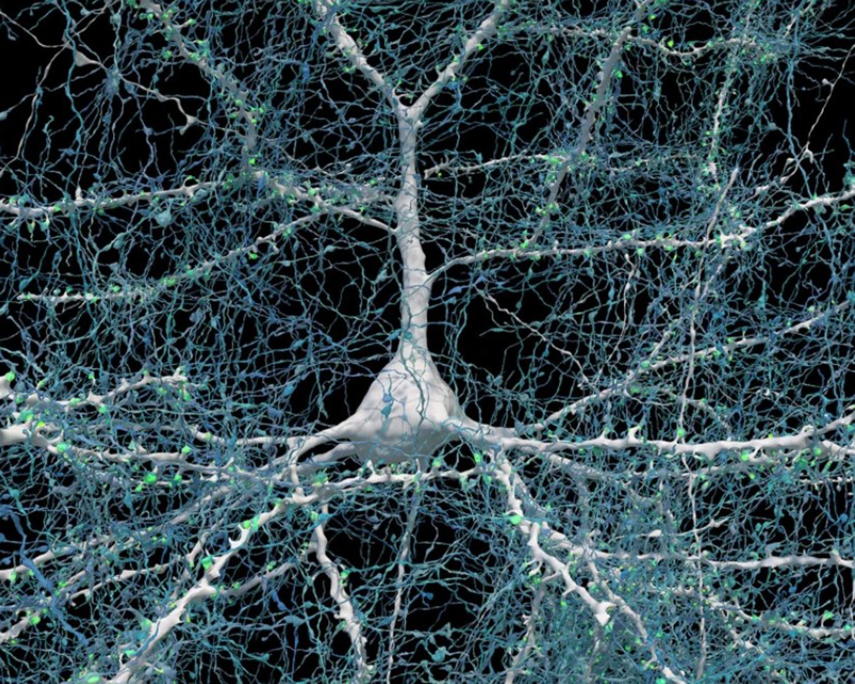
The problem with scientific research is that it never stops. Recently, my friend Jean-Pierre, who keeps a close eye on new articles in major neuroscientific journals, let me know about a major paper just published in Science, about how electron microscopy is now being used to reconstruct neurons of the human cortex with unequalled precision. And since this research had been done by Jeff Lichtman’s team at Harvard, and I had already written a post about it in this blog back in 2014 , I figured that I had better write another post about it now, because the images of neurons that this team has succeeded in producing recently are truly spectacular. For example, the image above represents one neuron with 5600 axons (blue) making synaptic connections (green)).
I’ll leave you here with a quote from the page where Lichtman and his team present their “browsable petascale reconstruction of the human cortex”, along with a few of their unbelievably precise images of some of the infinitesmally tiny structures inside our brains.
The Lichtman laboratory at Harvard University and the Connectomics at Google team are releasing the “H01” dataset and companion paper. H01 is a 1.4 petabyte volume of a small sample of human brain tissue. The sample was imaged at nanoscale-resolution by serial section electron microscopy, reconstructed and annotated by automated computational techniques, and analyzed for preliminary insights into the structure of human cortex.
The dataset comprises roughly one cubic millimeter of imaging data, including tens of thousands of reconstructed neurons, millions of neuron fragments, 183 million annotated synapses, 100 proofread cells [proofread by humans, because AI software sometimes makes errors in recognizing structures], and many additional subcellular annotations and structures — all easily accessible with the Neuroglancer browser interface.“
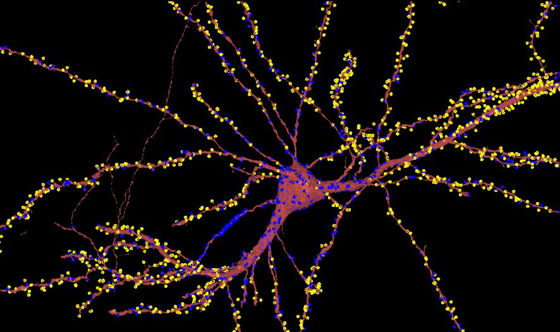
Excitatory synapses (yellow) and inhibitory synapses (blue) represented as points on the neuron that receive these connections.
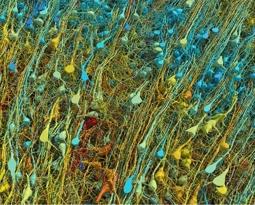
Rendering based on electron-microscopy data, showing the positions of the neurons in a fragment of the cerebral cortex. Neurons are colour-coded according to their size.
From the Simple to the Complex | Comments Closed