Tuesday, 1 May 2018
Limitations of Brain Mapping
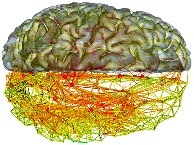
 With the use of tracers, dyes and modern brain-imaging technology, it is now possible to produce maps of the human brain. But why would we want to do so? Simply because our cognitive functions emerge from interactions among numerous parts of the brain, in many cases physically quite distant from one another. If we can map the neural highways that form the various networks in our brains, or better still, understand which are the preferred highways for use in particular situations, we can learn a lot about how we go about talking, thinking and behaving.
But because the brain is so complex, brain mapping involves many problems and is subject to some major limitations. In this post I will discuss just two of these problems and a few limitations inherent in efforts to map the “connectome”—to prepare a map of all the nerve bundles connecting the neurons to one another in a given brain.
The first problem concerns the spatial scale of the human brain. Simply stated, it is impossible to see all of the brain at any given moment in time. To illustrate this problem when I am giving a lecture, I often hold my two fists next to each other and ask the audience, if one represents a terminal of an axon in the brain and the other a dendritic spine on the other side of the synapse, how big would the entire brain be in proportion? The answer is about 40 km in diameter, roughly the length of Montreal Island. This means that if you’re using an electron microscope to map the brain on the micro scale of the synapses, as Sebastian Seung and Jeff Lichtman have done, you cannot simultaneously observe the areas of the brain from which the axons that terminate in these synapses originate.
Conversely, methods such as diffusion imaging and projects such as Big Brain let us see how large bundles of axonal nerve fibres spread out across the entire brain. But the resolution of these methods is too low to show the details of the synapses where these fibres connect to one another. That leaves the intermediate or meso scale, on which we can trace any path of the axons of particular neurons in the rat or mouse brain. But here again, we can cannot see the details of the synapses, and we are no longer dealing with the human brain.
The other major problem with brain mapping concerns the time scale involved. To be clear, we are not now and never will be able to prepare a definitive map of the connectome of a human brain, because the brain itself is extremely plastic. You could almost say that your brain’s primary activity consists in changing itself constantly, every hour, minute and even second of your life! So even if some day an imaging device were developed that was powerful enough to produce an image of your entire brain, that would be only one image of your brain at one moment in time. Just a few seconds later, your brain’s wiring would already have altered irretrievably at least a little bit.
In any case, at present science is still far from having established anything even approaching the entire human connectome. The only animal for which we have a complete map of the connectome is the small nematode (roundworm) C. elegans which measures 1 mm in length. Hence scientists have been able to produce a complete diagram of the connections among its 302 neurons, which form about 7000 synapses. But does that by definition mean that we now know everything about the behaviour of this little worm? Not a bit. Some authors even say that the entire effort has not really taught us much at all. But others argue that it has already enabled us to formulate numerous experimental hypotheses and test them successfully. In short, this is a subject on which there is much debate, as reported, for example, in the article “The Connectome Debate: Is Mapping the Mind of a Worm Worth It?”.
One of the points debated is that the “synaptic weight” of the connections is not known, It is all well and good to know which neuron connects to which other neuron, but not all connections are equally strong. Some synapses may have been strengthened through learning, while others have not, and so on. And there we come back to the problem of the brain’s changing over time.
We also do not know which of all these connections are excitatory and which are inhibitory. And that’s without even considering certain neuromodulators that circulate around the neurons and may change the way that they interact.
From the Simple to the Complex | No comments


