Monday, 21 November 2022
Zebrafish brain images may reveal neuronal bases of emotional memory
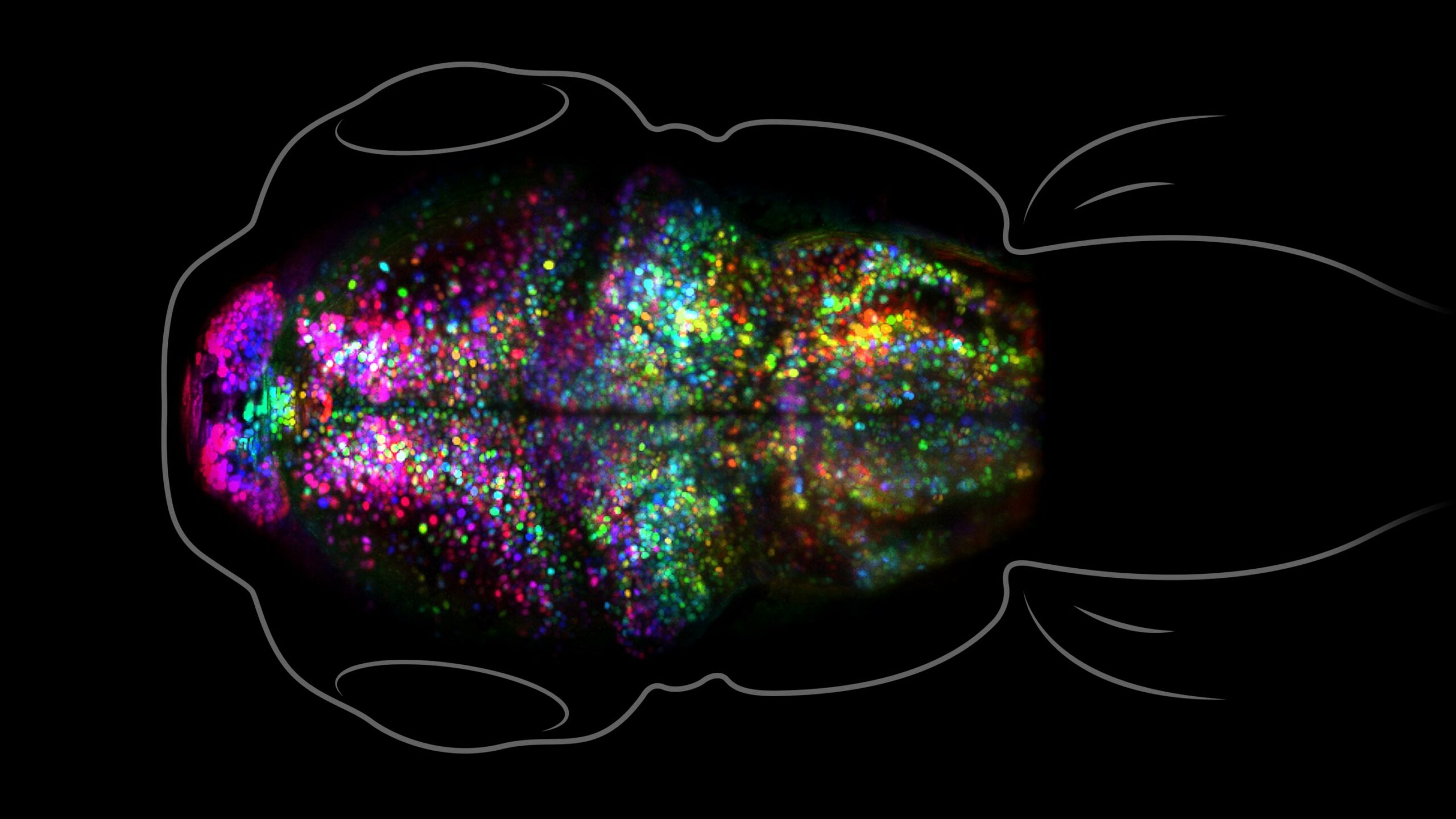
 As is well known, memories with heavy emotional connotations—especially negative ones—are very strong. In extreme cases of post-traumatic stress, such memories can be so recurrent and intrusive that they make life a living hell. In mammals, the brain structure most highly involved in such negative memories is the amygdala. But while much research has been done on the hippocampus, which is the brain structure involved in spatial, lexical and other forms of memory, the amygdala has received much less attention, in part because it is so hard to access. To get around this problem, a research team at the University of Southern California, in Los Angeles, used larval zebrafish and a fluorescence-based imaging method to visualize the changes that occurred in the synapses of the pallium (the fish brain structure equivalent to the amygdala) after aversive conditioning. The surprising results were published in the journal PNAS in January 2022, in an article entitled “Regional synapse gain and loss accompany memory formation in larval zebrafish”.
In the team’s experiments, the larval zebrafish were exposed to light together with heat to which they showed an aversive response, and some of them subsequently learned to respond to the light alone the same way that they had to the combination of light and heat. Before and after images were captured of the synapses in the pallium. Increases in the intensity of the fluorescence detected in these synapses would have indicated strengthening of these synapses, the most common mechanism for making neural circuits more efficient for a certain time (for example, in the hippocampus, for forming memories of new paths or new words). But surprisingly, the zebrafish who learned this conditioned response did not show any such increases in synaptic strength. Instead, they showed increases in the number of synapses in one part of the pallium and decreases in another—a far more radical mechanism than a mere increase in synaptic efficiency. The authors suggest that the explanation for this finding may lie in the more profound and durable nature of negative emotional conditioning, often associated with trauma.
Since its publication, however, this study has been criticized for having used larval-stage zebrafish rather than adults (this decision was made for practical reasons: unlike adults, larval zebrafish are still transparent, which makes their brain structures easier to access and visualize). In fish, just as in humans and other mammals, a great deal of synaptic pruning occurs naturally in early stages of development, so that only the synapses that get used the most survive. Hence, the critics argue, the pruning observed in this negative-conditioning study may not have been specific to this kind of learning, but simply to the natural process of synaptic pruning in the developing brain. As the saying goes, further research will be needed.
Memory and the Brain | Comments Closed


